
Syracuse University’s First NIH S10 Grant Funds the Purchase of State-of-the-Art Microscope
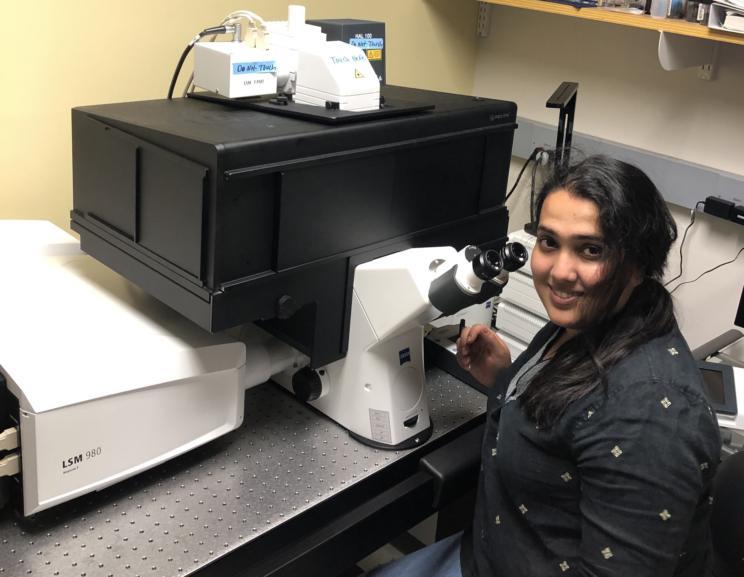
The Blatt BioImaging Center’s Zeiss LSM980 allows researchers to image their most challenging samples.
For the first time in Syracuse University’s history, a department has received a prestigious S10 Instrumentation grant from the National Institutes of Health. The S10 program, which supports the purchase of high-tech instruments to enhance research of NIH investigators, funded the acquisition of a Zeiss LSM980 confocal microscope. The new equipment will provide researchers at Syracuse University with sharp, detailed images of specific sections of cells.
The S10 grant was awarded to George Langford, A&S dean emeritus and professor emeritus of biology, in collaboration with colleagues at Syracuse University and SUNY Upstate.
Housed in the Department of Biology’s Blatt BioImaging Center, which is under the direction of assistant professor Heidi Hehnly, the confocal microscope adds to the Center’s variety of sensitive instruments. Thanks to many specialized microscopes, researchers have the equipment to investigate a wide range of samples, from microscopic level yeast cells all the way up to whole organisms. The Blatt BioImaging Center’s equipment is available for use with a reservation to anyone at Syracuse University, SUNY Upstate, SUNY ESF and other local institutions.
We recently caught up with Blatt BioImaging Center manager Mike Bates to learn more about the new confocal microscope and how it will benefit A&S faculty and student researchers.
How does a confocal microscope work?
The state-of-the-art Zeiss LSM980 allows us to view visual sections of tiny structures – such as embryos – that would be difficult to physically section, and construct 3D structures from the obtained images. The basic technique scans an object point-by-point using a focused laser beam to allow for a 3D reconstruction.
For someone not familiar with this technology, how is a confocal microscope different from other microscopes?
In a typical (“widefield”) microscope, the entire object is illuminated, but that can create blur from areas out of focus above and below the image plane. A confocal microscope scans a sample with a focused beam of light. The disadvantage of a widefield system is that it does not work well with thick samples that scatter light. For thicker samples, the confocal will give superior improvement in resolution. If a scientist is looking for a quick and dirty reason to choose one microscope over the other, they should consider if they have a thin or thick sample. A thin sample usually means widefield, tissues usually mean confocal.
What type of research are you currently using this microscope for?
The Hehnly lab uses the Zeiss 980 for increased resolution of intracellular events that occur during embryonic development, but a wide variety of research programs are using the Blatt BioImaging Center. We have seen researchers from disciplines including biology, chemistry, physics, forensics and engineering. These microscopes have been used to observe events in developmental and cellular biology, identify biophysical properties of developing tissues, and understanding protein-protein interactions.
Will students be able to use this technology?
Students can and are using the system currently. We actually encourage students who think they can benefit from the use of a confocal microscope or a widefield scope to bring their samples down and try some imaging.
What is the benefit to them in learning how to use a confocal microscope?
Learning microscopy adds a valuable skill to any student’s tool set because it will enable them to answer questions that may be of particular interest to researchers. Learning how to operate cutting-edge equipment such as the Zeiss 980 will pay dividends throughout their scientific career.
If someone is interested in using a microscope in the Blatt BioImaging Center, what should they do?
Contact myself, Mike Bates (mbates@syr.edu), or Heidi Hehnly (hhehnly@syr.edu). Also, check out the Blatt BioImaging Center for more details.